Brain Tumor Diagnosis
Clarity and Confidence
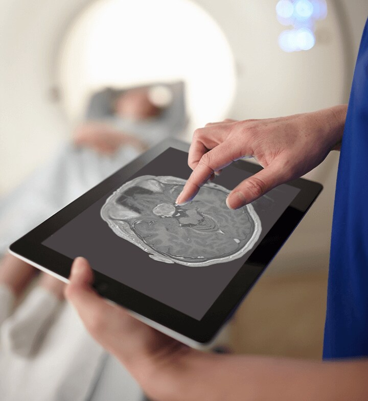
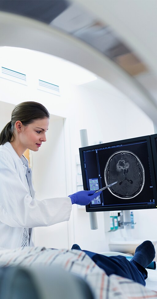
In brain tumor diagnosis Gadovist® can show comparably higher contrast enhancement than other macrocyclic gadolinium-based contrast agents (GBCAs).4,5
Studies in patients and preclinical studies in vivo4,5 show that in CNS imaging this:
• Delivers favorable image quality.
• Makes assessment of malignancy easier by providing higher contrast enhancement as well as higher SNR and CNR.
Higher contrast enhancement can lead to fast and more consistent diagnosis, which in turn can mean more effective treatment, to optimize the patient journey.


Multiple Sclerosis
Monitoring MS with Gadovist®
With gadolinium enhancement of active MS lesions blood-brain barrier (BBB) dysfunction can be shown.6,7 It is therefore a well-established marker of MS lesion inflammation.8 Using Gadovist®-enhanced Susceptibility-Weighted Imaging (SWI) has been shown to detect more active MS lesions than gadolinium T1 SE.7
MRI Techniques
Getting the Best from Gadovist®
Whether for brain tumor diagnosis, anatomic delineation, and treatment response, or for multiple sclerosis diagnosis, various MR imaging techniques can be chosen. Each has intrinsically different characteristics and can optimize the enhancement effects of Gadovist®.


New Dosing Option
Flexibility with Gadovist® in CNS Imaging
Gadovist® now offers more flexibility with a new dosing option. A label change triggered by the LEADER-75* study allows healthcare professionals to tailor procedures using a lower dose where they deem fit. This flexibility for dose reduction would be particularly relevant for patients who require multiple contrast-enhanced MRI examinations.9
The results of the study demonstrated the effectiveness of a lower dose of Gadovist® for CNS imaging. Gadovist® at a reduced dose of 0.075 mmol/kg body weight (BW) Gadovist® demonstrated a non-inferiority to the standard dose (0.1 mmol/kg BW) of gadoterate.**
Find out more by viewing our Q&A video, in which our medical scientific adviser answers the most asked questions in relation to the LEADER-75 study.
- * LEADER-75 (Lower Administered Dose With Higher Relaxivity: Gadovist® vs Dotarem®) Return to content
- ** at least 80% of the improvement over unenhanced imaging for all three efficacy variables was retained Return to content
References
- 1. Rohrer, Martin, et al. "Comparison of magnetic properties of MRI contrast media solutions at different magnetic field strengths." Investigative Radiology 40.11 (2005): 715-724. Return to content
- 2. Tombach, Bernd, et al. "Comparison of 1.0 M gadobutrol and 0.5 M gadopentate dimeglumine-enhanced MRI in 471 patients with known or suspected renal lesions: results of a multicenter, single-blind, interindividual, randomized clinical phase III trial." European Radiology 18.11 (2008): 2610-2619. Return to content
- 3. Tombach, Bernd, and Walter Heindel. "Value of 1.0-M gadolinium chelates: review of preclinical and clinical data on gadobutrol." European Radiology 12.6 (2002): 1550-1556. Return to content
- 4. Attenberger, Ulrike I., et al. "Evaluation of gadobutrol, a macrocyclic, nonionic gadolinium chelate in a brain glioma model: comparison with gadoterate meglumine and gadopentetate dimeglumine at 1.5 T, combined with an assessment of field strength dependence, specifically 1.5 versus 3 T." Journal of Magnetic Resonance Imaging: An Official Journal of the International Society for Magnetic Resonance in Medicine 31.3 (2010): 549-555. Return to content
- 5. Koenig, M., et al. "Intra-individual, randomised comparison of the MRI contrast agents gadobutrol versus gadoteridol in patients with primary and secondary brain tumours, evaluated in a blinded read." European Radiology 23.12 (2013): 3287-3295. Return to content
- 6. CM Poser, VV Brinar. "The nature of multiple sclerosis." Clin Neurol Neurosurg 106.3 (2004): 159-171. Return to content
- 7. LLF do Amaral, et al. "Gadolinium-enhanced susceptibility-weighted imaging in multiple sclerosis: optimizing the recognition of active plaques for different MR imaging sequences." AJNR Am J Neuroradiol 40.4 (2019): 614-619. Return to content
- 8. AG Kermode, et al. " Breakdown of the blood-brain barrier precedes symptoms and other MRI signs of new lesions in multiple sclerosis. Pathogenetic and clinical implications." Brain 113.(part 5) (1990): 1477-1489. Return to content
- 9. Liu BP, Rosenberg M, Saverio P, et al. Clinical Efficacy of Reduced-Dose Gadobutrol Versus Standard-Dose Gadoterate for Contrast-Enhanced MRI of the CNS: An International Multicenter Prospective Crossover Trial (LEADER-75). AJR Am J Roentgenol. 2021;217(5):1195-1205. doi:10.2214/AJR.21.25924 Return to content


